

| Yoon-Seok Chung | 5 Articles |
Objectives
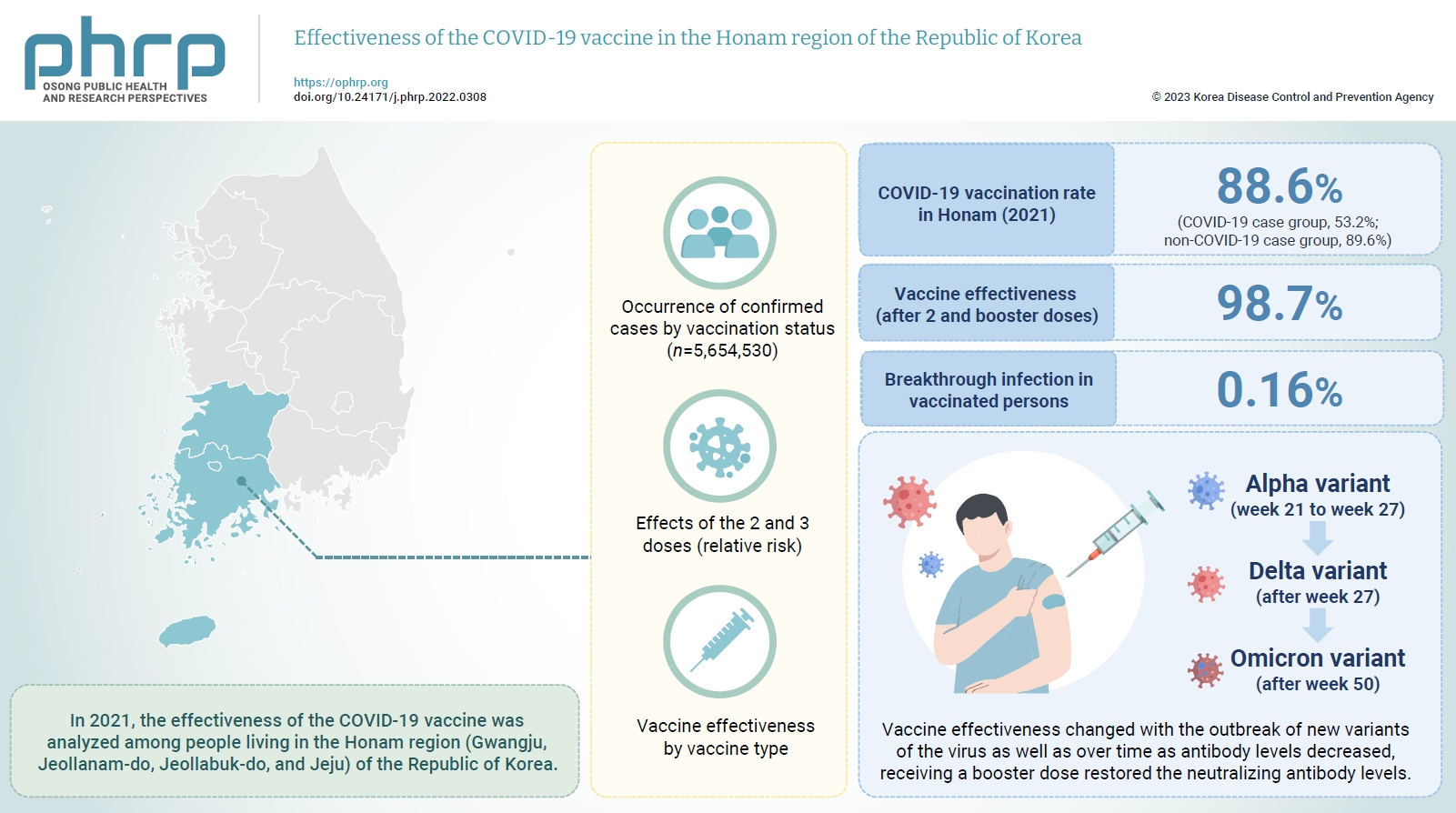
In 2021, the effectiveness of the COVID-19 vaccine was analyzed among people living in the Honam region (Gwangju, Jeollanam-do, Jeollabuk-do, and Jeju) of the Republic of Korea. And we investigated changes in the dominant virus strain. Methods This study used the data provided by the Korean Ministry of the Interior and Safety for individuals ≥12 years old in the Honam region, and the Integrated Disease and Health Management System of the Korea Centers for Disease Control and Prevention for COVID-19-vaccinated individuals as of December 31, 2021. Statistical analyzes were performed using IBM SPSS ver. 23.0. The occurrence of confirmed cases by vaccination status, the relative risk, and vaccine effectiveness by vaccine type were calculated. Results In 2021, the COVID-19 vaccination rate in Honam was 88.6%. The overall vaccine effectiveness (after 2 and 3 doses) was 98.7% (p<0.001). and the breakthrough infection rate was 0.16%. From week 21 to week 27 of 2021 (June 27 to July 3), the genome sequencing results were mostly alpha variants. The Delta variant emerged as the dominant variant after 27 weeks and the Omicron variant was found at 50 weeks (December 5–11). Conclusion Vaccine effectiveness changed with the outbreak of new variants of the virus as well as over time as antibody levels decreased. that the prevention effectiveness of vaccination in Honam was >98%, and the effect among persons who received 2 doses was >90% regardless of the vaccine type. Although vaccine effectiveness decreased because of reduced antibody levels over time (as observed in breakthrough infections), receiving a booster dose restored the neutralizing antibody levels.
<sec>
<title>Objectives</title>
<p>An epidemiological investigation was conducted into a hepatitis C virus (HCV) outbreak at an outpatients clinic in Seoul (2011–2012). The aim of the study was to analyze the scale of infection, identify the source of infection, and route of transmission to prevent hepatitis C transmission in the future.</p></sec>
<sec>
<title>Methods</title>
<p>A retrospective study of the outpatients and health care workers (<italic>n</italic> = 7,285) in the target outpatient clinic during 2011–2012 was conducted. The history of the study population infection with hepatitis C, electronic medical records, field visits, and health care worker interviews were examined for the period between March 1<sup>st</sup>, 2006 and March 25<sup>th</sup>, 2016. The blood samples were collected and tested for anti-HCV antibodies, HCV RNA and HCV gene in 2016.</p></sec>
<sec>
<title>Results</title>
<p>The rate of anti-HCV positive results was 4.4% in the study population. The risk factors associated with an anti-HCV positive result were ≥ 10 clinic visits, and receiving an invasive procedure including a nerve block and a block of the peripheral branch of the spinal nerve (<italic>p</italic> < 0.05). There were 112 HCV RNA positive cases out of 320 anti-HCV positive test result cases, amongst which 100 cases had the dominant HCV genotype 2a which formed either 1 cluster (<italic>n</italic> = 56) or 2 clusters (<italic>n</italic> = 25). This result indicated exposure to a high-association infection source.</p></sec>
<sec>
<title>Conclusion</title>
<p>Anti-HCV antibodies and genotypic analysis showed an epidemiological association between the outbreak of HCV and invasive procedures performed (2011–2012) at an outpatients clinic in Seoul.</p></sec>
<sec><title>Objectives</title>
<p>Coronavirus Disease-19 (COVID-19) is a respiratory infection characterized by the main symptoms of pneumonia and fever. It is caused by the novel coronavirus severe acute respiratory syndrome Coronavirus-2 (SARS-CoV-2), which is known to spread via respiratory droplets. We aimed to determine the rate and likelihood of SARS-CoV-2 transmission from COVID-19 patients through non-respiratory routes.</p></sec>
<sec><title>Methods</title>
<p>Serum, urine, and stool samples were collected from 74 hospitalized patients diagnosed with COVID-19 based on the detection of SARS-CoV-2 in respiratory samples. The SARS-CoV-2 RNA genome was extracted from each specimen and real-time reverse transcription polymerase chain reaction performed. CaCo-2 cells were inoculated with the specimens containing the SARS-COV-2 genome, and subcultured for virus isolation. After culturing, viral replication in the cell supernatant was assessed.</p></sec>
<sec><title>Results</title>
<p>Of the samples collected from 74 COVID-19 patients, SARS-CoV-2 was detected in 15 serum, urine, or stool samples. The virus detection rate in the serum, urine, and stool samples were 2.8% (9/323), 0.8% (2/247), and 10.1% (13/129), and the mean viral load was 1,210 ± 1,861, 79 ± 30, and 3,176 ± 7,208 copy/µL, respectively. However, the SARS-CoV-2 was not isolated by the culture method from the samples that tested positive for the SARS-CoV-2 gene.</p></sec>
<sec><title>Conclusion</title>
<p>While the virus remained detectable in the respiratory samples of COVID-19 patients for several days after hospitalization, its detection in the serum, urine, and stool samples was intermittent. Since the virus could not be isolated from the SARS-COV-2-positive samples, the risk of viral transmission via stool and urine is expected to be low.</p></sec>
Citations
Citations to this article as recorded by

<sec><title>Objectives</title>
<p>Severe acute respiratory syndrome coronavirus 2 (SARS-CoV-2) emerged in Wuhan, China, in December 2019 and has been rapidly spreading worldwide. Although the causal relationship among mutations and the features of SARS-CoV-2 such as rapid transmission, pathogenicity, and tropism, remains unclear, our results of genomic mutations in SARS-CoV-2 may help to interpret the interaction between genomic characterization in SARS-CoV-2 and infectivity with the host.</p></sec>
<sec><title>Methods</title>
<p>A total of 4,254 genomic sequences of SARS-CoV-2 were collected from the Global Initiative on Sharing all Influenza Data (GISAID). Multiple sequence alignment for phylogenetic analysis and comparative genomic approach for mutation analysis were conducted using Molecular Evolutionary Genetics Analysis (MEGA), and an in-house program based on Perl language, respectively.</p></sec>
<sec><title>Results</title>
<p>Phylogenetic analysis of SARS-CoV-2 strains indicated that there were 3 major clades including S, V, and G, and 2 subclades (G.1 and G.2). There were 767 types of synonymous and 1,352 types of non-synonymous mutation. ORF1a, ORF1b, S, and N genes were detected at high frequency, whereas ORF7b and E genes exhibited low frequency. In the receptor-binding domain (RBD) of the S gene, 11 non-synonymous mutations were observed in the region adjacent to the angiotensin converting enzyme 2 (ACE2) binding site.</p></sec>
<sec><title>Conclusion</title>
<p>It has been reported that the rapid infectivity and transmission of SARS-CoV-2 associated with host receptor affinity are derived from several mutations in its genes. Without these genetic mutations to enhance evolutionary adaptation, species recognition, host receptor affinity, and pathogenicity, it would not survive. It is expected that our results could provide an important clue in understanding the genomic characteristics of SARS-CoV-2.</p></sec>
Citations
Citations to this article as recorded by
<sec>
<title>Objectives</title>
<p>Following reports of patients with unexplained pneumonia at the end of December 2019 in Wuhan, China, the causative agent was identified as coronavirus (SARS-CoV-2), and the 2019 novel coronavirus disease was named COVID-19 by the World Health Organization. Putative patients with COVID-19 have been identified in South Korea, and attempts have been made to isolate the pathogen from these patients.</p></sec>
<sec>
<title>Methods</title>
<p>Upper and lower respiratory tract secretion samples from putative patients with COVID-19 were inoculated onto cells to isolate the virus. Full genome sequencing and electron microscopy were used to identify the virus.</p></sec>
<sec>
<title>Results</title>
<p>The virus replicated in Vero cells and cytopathic effects were observed. Full genome sequencing showed that the virus genome exhibited sequence homology of more than 99.9% with SARS-CoV-2 which was isolated from patients from other countries, for instance China. Sequence homology of SARS-CoV-2 with SARS-CoV, and MERS-CoV was 77.5% and 50%, respectively. Coronavirus-specific morphology was observed by electron microscopy in virus-infected Vero cells.</p></sec>
<sec>
<title>Conclusion</title>
<p>SARS-CoV-2 was isolated from putative patients with unexplained pneumonia and intermittent coughing and fever. The isolated virus was named BetaCoV/Korea/KCDC03/2020.</p></sec>
Citations
Citations to this article as recorded by
|
|


