

| Myung-Guk Han | 8 Articles |
Objectives
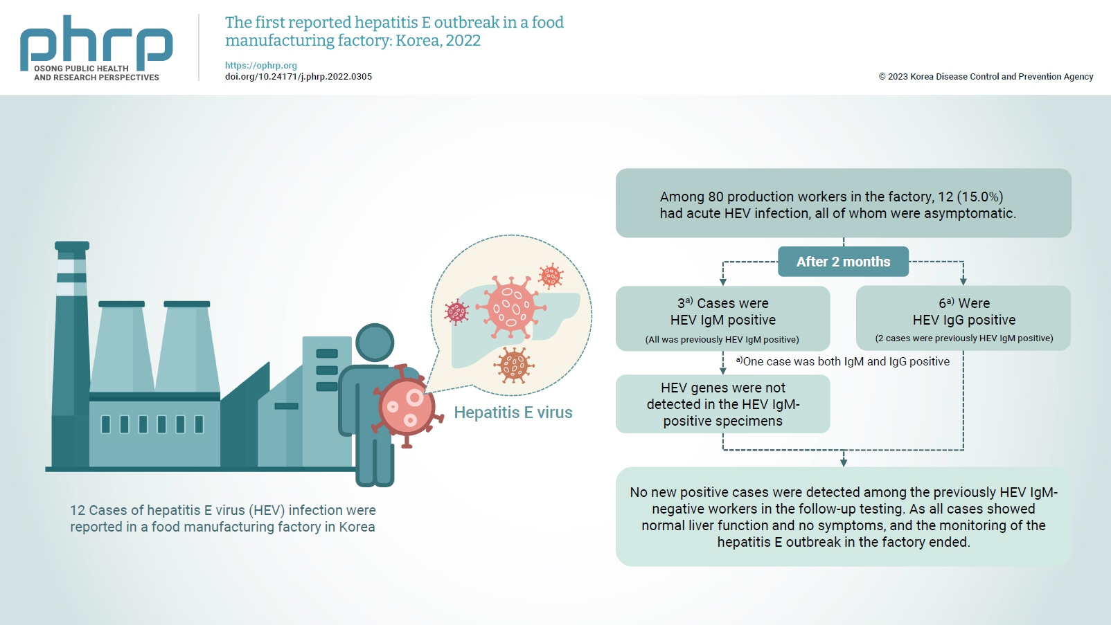
On February 16, 2022, 12 cases of hepatitis E virus (HEV) infection were reported in a food manufacturing factory in Korea. The aim of this study was to identify additional cases and to determine the source of this HEV outbreak. Methods: This study was an in-depth investigation of 12 HEV immunoglobulin M (IgM)-positive cases and their demographic, clinical, and epidemiological characteristics. On-site specimens were collected from the environment and from humans, and a follow-up investigation was conducted 2 to 3 months after the outbreak. Results: Among 80 production workers in the factory, 12 (15.0%) had acute HEV infection, all of whom were asymptomatic. The follow-up investigation showed that 3 cases were HEV IgMpositive, while 6 were HEV IgG-positive. HEV genes were not detected in the HEV IgM-positive specimens. HEV genes were not detected in the food products or environmental specimens collected on-site. HEV was presumed to be the causative pathogen. However, it could not be confirmed that the source of infection was common consumption inside the factory. Conclusion: This was the first domestic case of an HEV infection outbreak in a food manufacturing factory in Korea. Our results provide information for the future control of outbreaks and for the preparation of measures to prevent domestic outbreaks of HEV infection. Citations
Citations to this article as recorded by

The rotavirus vaccine is a live vaccine, and there is a possibility of infection by the virus strain used in the vaccine. We investigated the process of determining whether an infection was caused by the vaccine strain in a severe complex immunodeficiency (SCID) patient with rotavirus infection. The patient was vaccinated with RotaTeq prior to being diagnosed with SCID. The testing process was conducted in the following order: confirming rotavirus infection, determining its genotype, and confirming the vaccine strain. Rotavirus infection was confirmed through enzyme immunoassay and VP6 gene detection. G1 and P[8] were identified by multiplex polymerase chain reaction for the genotype, and G3 was further identified using a single primer. By detecting the fingerprint gene (WC3) of RotaTeq, it was confirmed that the detected virus was the vaccine strain. Genotypes G1 and P[8] were identified, and the infection was suspected of having been caused by rotavirus G1P[8]. G1P[8] is the most commonly detected genotype worldwide and is not included in the recombinant strains used in vaccines. Therefore, the infection was confirmed to have been caused by the vaccine strain by analyzing the genetic relationship between VP4 and VP7. Rotavirus infection by the vaccine strain can be identified through genotyping and fingerprint gene detection. However, genetic linkage analysis will also help to identify vaccine strains.
Citations
Citations to this article as recorded by
<p>Since a novel beta-coronavirus, severe acute respiratory syndrome coronavirus 2 (SARS-CoV-2) was first reported in December 2019, there has been a rapid global spread of the virus. Genomic surveillance was conducted on samples isolated from infected individuals to monitor the spread of genetic variants of SARS-CoV-2 in Korea. The Korea Disease Control and Prevention Agency performed whole genome sequencing of SARS-CoV-2 in Korea for 1 year (January 2020 to January 2021). A total of 2,488 SARS-CoV-2 cases were sequenced (including 648 cases from abroad). Initially, the prevalent clades of SARS-CoV-2 were the S and V clades, however, by March 2020, GH clade was the most dominant. Only international travelers were identified as having G or GR clades, and since the first variant 501Y.V1 was identified (from a traveler from the United Kingdom on December 22<sup>nd</sup>, 2020), a total of 27 variants of 501Y.V1, 501Y.V2, and 484K.V2 have been classified (as of January 25<sup>th</sup>, 2021). The results in this study indicated that quarantining of travelers entering Korea successfully prevented dissemination of the SARS-CoV-2 variants in Korea.</p>
Citations
Citations to this article as recorded by
<sec>
<title>Objectives</title>
<p>The Korea Centers for Disease Control and Prevention has published “A Guideline for Unknown Disease Outbreaks (UDO).” The aim of this report was to introduce tabletop exercises (TTX) to prepare for UDO in the future.</p></sec>
<sec>
<title>Methods</title>
<p>The UDO Laboratory Analyses Task Force in Korea Centers for Disease Control and Prevention in April 2018, assigned unknown diseases into 5 syndromes, designed an algorithm for diagnosis, and made a panel list for diagnosis by exclusion. Using the guidelines and laboratory analyses for UDO, TTX were introduced.</p></sec>
<sec>
<title>Results</title>
<p>Since September 9<sup>th</sup>, 2018, the UDO Laboratory Analyses Task Force has been preparing TTX based on a scenario of an outbreak caused by a novel coronavirus. In December 2019, through TTX, individual missions, epidemiological investigations, sample treatments, diagnosis by exclusions, and next generation sequencing analysis were discussed, and a novel coronavirus was identified as the causal pathogen.</p></sec>
<sec>
<title>Conclusion</title>
<p>Guideline and laboratory analyses for UDO successfully applied in TTX. Conclusions drawn from TTX could be applied effectively in the analyses for the initial response to COVID-19, an ongoing epidemic of 2019 – 2020. Therefore, TTX should continuously be conducted for the response and preparation against UDO.</p></sec>
Citations
Citations to this article as recorded by
<sec><title>Objectives</title>
<p>Coronavirus Disease-19 (COVID-19) is a respiratory infection characterized by the main symptoms of pneumonia and fever. It is caused by the novel coronavirus severe acute respiratory syndrome Coronavirus-2 (SARS-CoV-2), which is known to spread via respiratory droplets. We aimed to determine the rate and likelihood of SARS-CoV-2 transmission from COVID-19 patients through non-respiratory routes.</p></sec>
<sec><title>Methods</title>
<p>Serum, urine, and stool samples were collected from 74 hospitalized patients diagnosed with COVID-19 based on the detection of SARS-CoV-2 in respiratory samples. The SARS-CoV-2 RNA genome was extracted from each specimen and real-time reverse transcription polymerase chain reaction performed. CaCo-2 cells were inoculated with the specimens containing the SARS-COV-2 genome, and subcultured for virus isolation. After culturing, viral replication in the cell supernatant was assessed.</p></sec>
<sec><title>Results</title>
<p>Of the samples collected from 74 COVID-19 patients, SARS-CoV-2 was detected in 15 serum, urine, or stool samples. The virus detection rate in the serum, urine, and stool samples were 2.8% (9/323), 0.8% (2/247), and 10.1% (13/129), and the mean viral load was 1,210 ± 1,861, 79 ± 30, and 3,176 ± 7,208 copy/µL, respectively. However, the SARS-CoV-2 was not isolated by the culture method from the samples that tested positive for the SARS-CoV-2 gene.</p></sec>
<sec><title>Conclusion</title>
<p>While the virus remained detectable in the respiratory samples of COVID-19 patients for several days after hospitalization, its detection in the serum, urine, and stool samples was intermittent. Since the virus could not be isolated from the SARS-COV-2-positive samples, the risk of viral transmission via stool and urine is expected to be low.</p></sec>
Citations
Citations to this article as recorded by
<sec><title>Objectives</title>
<p>Severe acute respiratory syndrome coronavirus 2 (SARS-CoV-2) emerged in Wuhan, China, in December 2019 and has been rapidly spreading worldwide. Although the causal relationship among mutations and the features of SARS-CoV-2 such as rapid transmission, pathogenicity, and tropism, remains unclear, our results of genomic mutations in SARS-CoV-2 may help to interpret the interaction between genomic characterization in SARS-CoV-2 and infectivity with the host.</p></sec>
<sec><title>Methods</title>
<p>A total of 4,254 genomic sequences of SARS-CoV-2 were collected from the Global Initiative on Sharing all Influenza Data (GISAID). Multiple sequence alignment for phylogenetic analysis and comparative genomic approach for mutation analysis were conducted using Molecular Evolutionary Genetics Analysis (MEGA), and an in-house program based on Perl language, respectively.</p></sec>
<sec><title>Results</title>
<p>Phylogenetic analysis of SARS-CoV-2 strains indicated that there were 3 major clades including S, V, and G, and 2 subclades (G.1 and G.2). There were 767 types of synonymous and 1,352 types of non-synonymous mutation. ORF1a, ORF1b, S, and N genes were detected at high frequency, whereas ORF7b and E genes exhibited low frequency. In the receptor-binding domain (RBD) of the S gene, 11 non-synonymous mutations were observed in the region adjacent to the angiotensin converting enzyme 2 (ACE2) binding site.</p></sec>
<sec><title>Conclusion</title>
<p>It has been reported that the rapid infectivity and transmission of SARS-CoV-2 associated with host receptor affinity are derived from several mutations in its genes. Without these genetic mutations to enhance evolutionary adaptation, species recognition, host receptor affinity, and pathogenicity, it would not survive. It is expected that our results could provide an important clue in understanding the genomic characteristics of SARS-CoV-2.</p></sec>
Citations
Citations to this article as recorded by
<sec>
<title>Objectives</title>
<p>Following reports of patients with unexplained pneumonia at the end of December 2019 in Wuhan, China, the causative agent was identified as coronavirus (SARS-CoV-2), and the 2019 novel coronavirus disease was named COVID-19 by the World Health Organization. Putative patients with COVID-19 have been identified in South Korea, and attempts have been made to isolate the pathogen from these patients.</p></sec>
<sec>
<title>Methods</title>
<p>Upper and lower respiratory tract secretion samples from putative patients with COVID-19 were inoculated onto cells to isolate the virus. Full genome sequencing and electron microscopy were used to identify the virus.</p></sec>
<sec>
<title>Results</title>
<p>The virus replicated in Vero cells and cytopathic effects were observed. Full genome sequencing showed that the virus genome exhibited sequence homology of more than 99.9% with SARS-CoV-2 which was isolated from patients from other countries, for instance China. Sequence homology of SARS-CoV-2 with SARS-CoV, and MERS-CoV was 77.5% and 50%, respectively. Coronavirus-specific morphology was observed by electron microscopy in virus-infected Vero cells.</p></sec>
<sec>
<title>Conclusion</title>
<p>SARS-CoV-2 was isolated from putative patients with unexplained pneumonia and intermittent coughing and fever. The isolated virus was named BetaCoV/Korea/KCDC03/2020.</p></sec>
Citations
Citations to this article as recorded by
To compare the epidemiological characteristics of dengue cases imported by travelers or immigration in both Korea and Japan, we determined dengue incidence and related risk factors. During 2006–2010, 367 and 589 imported dengue cases were reported in Korea and Japan, respectively. In Korea, the presumptive origins for the dengue infections were Southeast Asia (82.6%), Southern Asia (13.9%), Eastern Asia (1.1%), South America (0.3%), Central America (0.3%), Africa (0.3%), and other countries (1.6%). In Japan, the origins of the infections were Southeast Asia (69.8%), Southern Asia (20.0%), Eastern Asia (1.7%), South America (2.5%), Central America (1.2%), Africa (1.2%), Oceania (2.4%), and other countries (1.2%). In both countries, more dengue cases were reported for men than for women (<i>p</i> < 0.01), and those aged 20–30 years accounted for > 60% of the total cases. The frequency of imported cases in summer and autumn (∼70% of total cases) was similar in both countries. This study demonstrates that there is a similar pattern of imported dengue cases in Korea and Japan. Therefore, there is a risk of an autochthonous dengue outbreak in Korea, as indicated by the recent outbreak in Japan in 2014.
Citations
Citations to this article as recorded by
|
|


