
Articles
- Page Path
- HOME > Osong Public Health Res Perspect > Volume 7(1); 2016 > Article
-
Original Article
Effects of Electromagnetic Radiation from Smartphones on Learning Ability and Hippocampal Progenitor Cell Proliferation in Mice - Yu-Jin Choi, Yun-Sik Choi
-
Osong Public Health and Research Perspectives 2015;7(1):12-17.
DOI: https://doi.org/10.1016/j.phrp.2015.12.009
Published online: December 23, 2015
Department of Pharmaceutical Science and Technology, Catholic University of Daegu, Gyeongsan, Korea
- ∗Corresponding author.
• Received: November 16, 2015 • Accepted: December 17, 2015
Copyright © 2015 Korea Centers for Disease Control and Prevention. Published by Elsevier Korea LLC.
This is an open access article under the CC BY-NC-ND license (http://creativecommons.org/licenses/by-nc-nd/4.0/).
Abstract
-
Objectives
- Nonionizing radiation is emitted from electronic devices, such as smartphones. In this study, we intended to elucidate the effect of electromagnetic radiation from smartphones on spatial working memory and progenitor cell proliferation in the hippocampus.
-
Methods
- Both male and female mice were randomly separated into two groups (radiated and control) and the radiated group was exposed to electromagnetic radiation for 9 weeks and 11 weeks for male and female mice, respectively. Spatial working memory was examined with a Y maze, and proliferation of hippocampal progenitor cells were examined by 5-bromo-2′-deoxyuridine administration and immunohistochemical detection.
-
Results
- When spatial working memory on a Y maze was examined in the 9th week, there was no significant difference in the spontaneous alternation score on the Y maze between the two groups. In addition, there was no significant difference in hippocampal progenitor cell proliferation. However, immunoreactivity to glial fibrillary acidic protein was increased in exposed animals. Next, to test the effect of recovery following chronic radiation exposure, the remaining female mice were further exposed to electromagnetic radiation for 2 more weeks (total 11 weeks), and spontaneous alternation was tested 4 weeks later. In this experiment, although there was no significant difference in the spontaneous alternation scores, the number of arm entry was significantly increased.
-
Conclusion
- These data indicate that although chronic electromagnetic radiation does not affect spatial working memory and hippocampal progenitor cell proliferation it can mediate astrocyte activation in the hippocampus and delayed hyperactivity-like behavior.
- Nonionizing electromagnetic radiation is energy that is given off from energy sources, including power lines, mobile phones, common electrical devices, and some types of machinery. This type of radiation differs from ionizing radiation, such as gamma rays, X-rays, and ultraviolet light, which exhibit high-frequency waves and have enough energy to liberate an electron from molecules [1]. Although nonionizing radiation has a lower frequency and is generally considered safe, accumulating evidence suggests that some types of nonionizing electromagnetic radiation have enough energy to harm living tissues. Especially, in recent years, the number of smartphone users has tremendously increased; thus, concerns and debates regarding the effects on human health of nonionizing radiation from smartphones have emerged. Importantly, unless some protective measures, such as hands-free or Bluetooth, are employed, the majority of people speak over smartphones with them near the users' heads. Therefore, it is an urgent issue to elucidate the effect of nonionizing radiation from smartphones or mobile phones on development and brain function.
- In this study, to elucidate the effect of chronic exposure to electromagnetic radiation from smartphones, we examined adult progenitor cell proliferation in the hippocampus, as well as special learning ability using a Y maze in mice.
Introduction
- 2.1 Animals
- Male and female C57BL/6 mice (8 wk old, Koatec, Kyoungki-do, Korea) were housed at a standard temperature (22 ± 1°C), humidity (50 ± 5%), and in a light-controlled environment (lights on from 8:00 am to 8:00 pm) with ad libitum access to food and water. The experimental protocol was approved by the Institutional Animal Care and Use Committee of the Catholic University of Daegu (IACUC-2012-34).
- 2.2 Exposure to electromagnetic radiation
- Mice were divided into two groups: animals from the exposed group were housed in a cage that was placed on the smartphone (Galaxy K, Samsung Electronics, South Korea) and it was maintained on operating mode throughout the study. To imitate the condition of daily smartphone use in life, we called and let mice hear the sound from the smartphone for 10 minutes a day. In the control group, animals were housed under normal conditions without electromagnetic radiation exposure. Mice were exposed to electromagnetic radiation for 9 weeks, and spontaneous alternation was tested. To verify the effect of recovery following electromagnetic radiation exposure, female mice were further exposed to electromagnetic radiation for 2 more weeks. After the smartphone was removed, female mice were housed under normal conditions for 4 weeks to provide a recovery period, and spontaneous alternation was tested (Figure 1).
- 2.3 Spontaneous alternation in a Y maze
- A Y maze was made from black Plexiglas and consisted of three arms with an angle of 120° between each arm. Spontaneous alternation consists of sequential entry into all three arms. Percent alternation was calculated by dividing the number of alternations by the number of possible alternations [number of alternation/(number of total arm entries − 2)].
- 2.4 Injection of 5-bromo-2′-deoxyuridine and immunohistochemistry
- To label proliferating cells, mice received an intraperitoneal (i.p.) injection of 5-bromo-2′-deoxyuridine (BrdU; 100 mg/kg, dissolved in saline; Sigma Aldrich, St. Louis, MO, USA) and were sacrificed 1 day later. A BrdU immunohistochemistry method was described by Choi et al [2]. To label glial fibrillary acidic protein (GFAP) and CD68, sections were blocked with 10% normal goat serum, followed by overnight incubation with a mouse monoclonal anti-GFAP antibody (1:1,000; Millipore, Temecula, CA, USA) or a monoclonal anti-CD68 antibody (1:500; Biolegend, San Diego, CA, USA). After several washes with phosphate buffered saline, sections were incubated (2 h at room temperature) with secondary antibodies conjugated with horseradish peroxidase (1:1,000; Jackson Immunoresearch, West Grove, PA, USA) and developed using diaminobenzidine (0.1%, Sigma Aldrich) and hydrogen proxidase (0.005%, Junsei Chemical, Tokyo, Japan).
- 2.5 Cell quantitation
- The density of BrdU-positive cells was quantitated as described by Choi et al [2]. Briefly, cells were counted (bilaterally) in three dorsal hippocampal sections (AP coordinate of the first dorsal-most section: –1.40 μm) separated by 160-μm intervals and averaged for each animal. To measure the granule cell layer/subgranular zone area, photomicrographs of sections were captured (10×) and quantitation was performed using Photoshop (Adobe Systems Incorporated, San Jose, CA, USA). The granule cell layers/subgranular zones of both the upper and lower blades were outlined and the area was measured. Volume was counted in three sections using the coordinates described above. The number of cells was expressed as the mean ± standard error of the mean from five mice for each group. Cell counts were analyzed statistically using the Student t test, and significance was accepted at p < 0.05.
Materials and methods
- 3.1 The effect of electromagnetic radiation on the Y maze task
- First, to elucidate the effect of nonionizing radiation from smartphones on spatial working memory, we measured spontaneous alternations using a Y maze. For this purpose, 4-week-old mice were randomly separated into two groups, and animals from one group were exposed to electromagnetic radiation emitted by a smartphone for 9 weeks (exposed group: 5 males and 8 females). The control group mice (5 males and 10 females) were housed under standard conditions without any electromagnetic radiation from the smartphone. In this experiment, the percent of alternation was not significantly different between the two groups (Figure 2A). In addition, there was no significant difference in arm entry (Figure 2B). These data indicate that chronic electromagnetic radiation exposure from smartphones for 9 weeks does not affect spatial working memory in mice.
- 3.2 The effect of electromagnetic radiation on progenitor cell proliferation in the dentate gyrus
- Next, to elucidate whether electromagnetic radiation emitted by a smartphone affects progenitor cell proliferation in the dentate gyrus, male mice from both groups were injected with BrdU (100 mg/kg) 1 day after the Y maze test and transcardially perfused 24 hours later. As shown in Figure 3, although the density of BrdU in the subgranular zone of the exposed group was slightly higher than that of the control animals, there was no significant difference between the two groups. These data also indicate that exposure to electromagnetic radiation for 9 weeks does not have any significant influence on progenitor cell proliferation in the subgranular zone.
- 3.3 The effect of electromagnetic radiation on glial reaction in the hippocampus
- Next, to identify whether chronic exposure to electromagnetic radiation has an effect on glial reaction immunoreactivity to GFAP, a marker of astrocytes, and CD68, a marker of microglia/macrophages, was examined. For this study, sections from the controls or from the mice exposed to electromagnetic radiation for 9 weeks were stained, and, as shown in Figure 4, increased immunoreactivity to GFAP was observed in the hippocampus, especially in the CA1 subfield. However, there was no difference in immunoreactivity to CD68 (data not shown).
- 3.4 The effect of recovery after electromagnetic radiation on the Y maze task
- Finally, we tested whether recovery following chronic exposure to electromagnetic radiation affects spatial working memory. For this purpose, female mice tested using the Y maze above were further exposed to electromagnetic radiation emitted from a smartphone for 2 more weeks (total of 11 weeks); then, smartphones were removed and mice were housed under normal conditions for 4 weeks to provide a recovery period. As shown in Figure 5, the percentage of spontaneous alternation was not significantly different compared with that measured after exposure to electromagnetic radiation for 9 weeks. However, the number of arm entries was significantly increased. These data indicate that chronic exposure to electromagnetic radiation may induce hyperactivity-like behavior later on.
Results
- At present, people are constrainedly exposed to the electromagnetic radiation given off from the electronic devices, including mobile phones and many types of machinery. As electromagnetic waves have been known to be capable of breaking chemical bonds and harming living tissues, extensive research has been focused on elucidating the impact of electromagnetic radiation on human health. However, in view of the results thus far achieved, it is quite controversial. For example, radiation from mobile phones or electromagnetic fields of generators increased the intracellular concentration of reactive oxygen species, activated apoptotic signaling, and induced marked cell death 3, 4, 5, 6, 7. On the other hand, exposure to electromagnetic fields does not alter apoptosis and even enhances progenitor cell proliferation and its survival in the hippocampus 8, 9, 10. In human studies, as the majority so far has been focused on human health following acute to subchronic exposure to electromagnetic radiation, it is not easy to translate results to the circumstance of smartphone use in real life. Of note is that many people carry smartphones in their pockets all day long and sleep with them next to them or even under the pillow. That being said, it is urgent to evaluate carefully the physiological effect of long-term exposure to the electromagnetic radiation from smartphones. To this end, male and female mice were exposed to the electromagnetic field for 9 weeks and 11 weeks, respectively, and spatial working memory and progenitor cell proliferation in the dentate gyrus were examined.
- To assess spatial working memory, we employed a spontaneous alternation test with a Y maze apparatus. In this experiment, exposure to the radiation field for 9 weeks did not significantly change the percentage of spontaneous alternation or the number of arm entries in both male and female animals. In addition, there was no significant difference in progenitor cell proliferation in the dentate gyrus. As adult neurogenesis in the dentate gyrus has been well known to play an important role in spatial working memory, these behavioral and physiological data indicate that chronic exposure to electromagnetic radiation does not affect spatial working memory in mice.
- Recently, it was reported that prenatal exposure to electromagnetic radiation resulted in hyperactivity in the open field test or the light-dark box test in rodents 11, 12. In addition, prenatal and, to a lesser degree, postnatal exposure to cell phones is associated with hyperactivity problems around the age of school entry [13]. These reports led us to test whether recovery after chronic electromagnetic radiation exposure affects spatial working memory and/or animal behavior. To answer these questions, we exposed electromagnetic radiation to female mice for 11 weeks and provided a recovery period under normal housing condition for 4 weeks. To our surprise, although the percentage of spontaneous alternation was not affected, the number of arm entries was significantly increased by providing a recovery for 4 weeks following an 11-week-long period of exposure to a radiation field. These data further support the previous evidence showing that exposure to electromagnetic radiation may be able to induce delayed hyperactivity-like behavior.
- In our study, there was no significant difference in progenitor cell proliferation in the hippocampus and in spatial working memory. However, some reports indicate that electromagnetic radiation influences progenitor cell proliferation and/or spatial working memory 14, 15. The reason for the discord is unknown, although it is worth noting that we provided exposure to electromagnetic radiation for a relatively long term. Interestingly, recent data indicate that although electromagnetic fields influence learning and memory in rodents, the animals can adapt to long-term exposure [16]. In addition, long-term whole-body exposure to an electromagnetic field from a mobile phone does not cause any adverse effects on memory function and development [17]. Therefore, it is tempting to hypothesize that our nervous system can adapt to long-term exposure to electromagnetic radiation from smartphones. However, we cannot exclude the possibility that acute whole-body exposure to electromagnetic radiation changes working memory and/or hippocampal progenitor cell proliferation. Further study will be needed to elucidate this hypothesis.
- In summary, we provided herein further solid evidence supporting the hypothesis that chronic exposure to electromagnetic radiation may induce delayed hyperactivity-like behavior without affecting spatial working memory and hippocampal progenitor cell proliferation.
Discussion
- The authors have no conflicts of interest to declare.
Conflicts of interest
-
Acknowledgements
- This work was supported by the Korean Foundation for the Advancement of Science & Creativity (HCR. 2013).
Acknowledgments
- 1. Genuis S.J.. Fielding a current idea: exploring the public health impact of electromagnetic radiation. Public Health 122(2). 2008 Feb;113−124. PMID: 17572456.ArticlePubMed
- 2. Choi Y.S., Karelina K., Alzate-Correa D.. Mitogen- and stress-activated kinases regulate progenitor cell proliferation and neuron development in the adult dentate gyrus. J Neurochem 123(5). 2012 Dec;676−688. PMID: 23020821.ArticlePubMed
- 3. Avci B., Akar A., Bilgici B.. Oxidative stress induced by 1.8 GHz radio frequency electromagnetic radiation and effects of garlic extract in rats. Int J Radiat Biol 88(11). 2012 Nov;799−805. PMID: 22788526.ArticlePubMed
- 4. Hou Q., Wang M., Wu S.. Oxidative changes and apoptosis induced by 1800-MHz electromagnetic radiation in NIH/3T3 cells. Electromagn Biol Med 34(1). 2015 Mar;85−92. PMID: 24665905.ArticlePubMed
- 5. Liu M.L., Wen J.Q., Fan Y.B.. Potential protection of green tea polyphenols against 1800 MHz electromagnetic radiation-induced injury on rat cortical neurons. Neurotox Res 20(3). 2011 Oct;270−276. PMID: 21293955.ArticlePubMed
- 6. Liu Y.X., Tai J.L., Li G.Q.. Exposure to 1950-MHz TD-SCDMA electromagnetic fields affects the apoptosis of astrocytes via caspase-3-dependent pathway. PLoS One 7(8). 2012 Aug;e42332PMID: 22870319.ArticlePubMed
- 7. Luo Y., Wang X., Chen Y.. Effects of electromagnetic radiation on morphology and TGF-β3 expression in mouse testicular tissue. Toxicology 310:2013 Aug;8−14. PMID: 23707491.ArticlePubMed
- 8. Cuccurazzu B., Leone L., Podda M.V.. Exposure to extremely low-frequency (50 Hz) electromagnetic fields enhances adult hippocampal neurogenesis in C57BL/6 mice. Exp Neurol 226(1). 2010 Nov;173−182. PMID: 20816824.ArticlePubMed
- 9. Misa Agustiño M.J., Leiro J.M., Jorge Mora M.T.. Electromagnetic fields at 2.45 GHz trigger changes in heat shock proteins 90 and 70 without altering apoptotic activity in rat thyroid gland. Biol Open 1(9). 2012 Sep;831−838. PMID: 23213477.ArticlePubMed
- 10. Podda M.V., Leone L., Barbati S.A.. Extremely low-frequency electromagnetic fields enhance the survival of newborn neurons in the mouse hippocampus. Eur J Neurosci 39(6). 2014 Mar;893−903. PMID: 24382162.ArticlePubMed
- 11. Wang H., Chen D., Gao C.. Effects of low level prenatal 60Co gamma-irradiation on postnatal growth and behavior in mice. Teratology 48(5). 1993 Nov;451−457. PMID: 8303614.ArticlePubMed
- 12. Aldad T.S., Gan G., Gao X.B.. Fetal radiofrequency radiation exposure from 800–1900 mHz-rated cellular telephones affects neurodevelopment and behavior in mice. Sci Rep 2:2012 Mar;312PMID: 22428084.ArticlePubMed
- 13. Divan H.A., Kheifets L., Obel C.. Prenatal and postnatal exposure to cell phone use and behavioral problems in children. Epidemiology 19(4). 2008 Jul;523−529. PMID: 18467962.ArticlePubMed
- 14. Fragopoulou A.F., Miltiadous P., Stamatakis A.. Whole body exposure with GSM 900 MHz affects spatial memory in mice. Pathophysiology 17(3). 2010 Jun;179−187. PMID: 19954937.ArticlePubMed
- 15. Ntzouni M.P., Stamatakis A., Stylianopoulou F.. Short-term memory in mice is affected by mobile phone radiation. Pathophysiology 18(3). 2011 Jun;193−199. PMID: 21112192.ArticlePubMed
- 16. Hao D., Yang L., Chen S.. Effects of long-term electromagnetic field exposure on spatial learning and memory in rats. Neurol Sci 34(2). 2013 Feb;157−164. PMID: 22362331.ArticlePubMed
- 17. Takahashi S., Imai N., Nabae K.. Lack of adverse effects of whole-body exposure to a mobile telecommunication electromagnetic field on the rat fetus. Radiat Res 173(3). 2010 Mar;362−372. PMID: 20199221.ArticlePubMed
References
Figure 1Summary of experimental schedule. Male and female mice were divided into control and exposed groups. Mice from the exposed group were exposed to electromagnetic radiation for 9 weeks and spatial working memory was measured. One hour after measurement 5-bromo-2′-deoxyuridine (BrdU) was injected into male mice from both groups and BrdU-injected animals were sacrificed 1 day later. Female mice were housed 2 more weeks under electromagnetic radiation or control conditions and housed for 4 more weeks without electromagnetic radiation to give the exposed group recovery. At the 15th week spatial working memory was measured again with a Y maze.
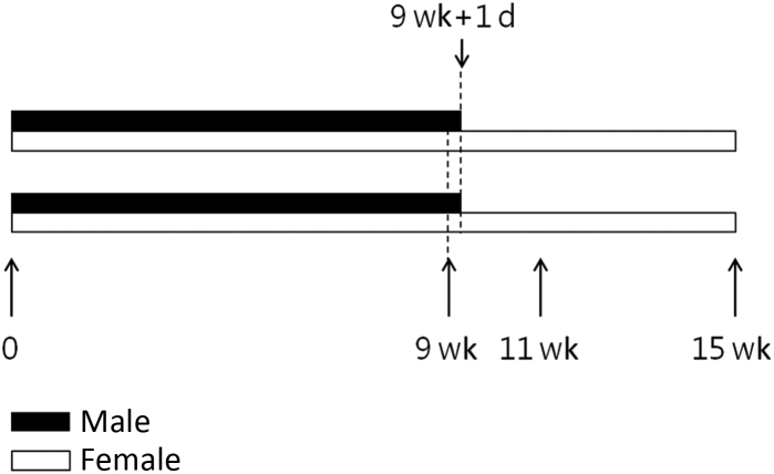
Figure 2Spatial working memory examined after electromagnetic exposure for 9 weeks. (A) Spontaneous alternation score was not significantly different between control and exposed groups. Data were collected from 5 males and 10 females from the control group, and 5 males and 8 females from the exposed group. (B) The number of arm entry was not significantly different between control and exposed groups. n.s. = not significant.
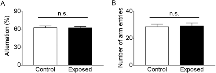
Figure 3Hippocampal progenitor cell proliferation. Hippocampal progenitor cell proliferation was monitored following intraperitoneal injection of 5-bromo-2′-deoxyuridine (BrdU). (A) Representative images show the expression pattern of BrdU in the subgranular zone of the hippocampus from control and exposed groups. (B) Mean density of BrdU-labeled cells were not significantly different between two groups suggesting that exposure to the electromagnetic radiation does not affect progenitor cell proliferation in the hippocampus. BrdU = 5-bromo-2′-deoxyuridine; con = control; exp = exposed; n.s. = not significant.
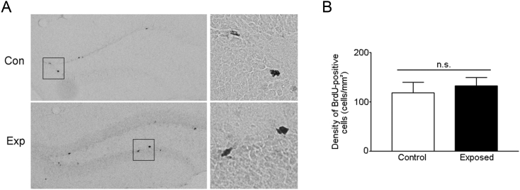
Figure 4Astrocyte activation by chronic electromagnetic radiation exposure. Compared to control (A), immunoreactivity to glial fibrillary acidic protein (GFAP), a marker of reactive astrocyte, increased in radiated animals (B). Among the hippocampal subregions GFAP immunoreactivity distinctively increased in CA1 area rather than dentate gyrus.
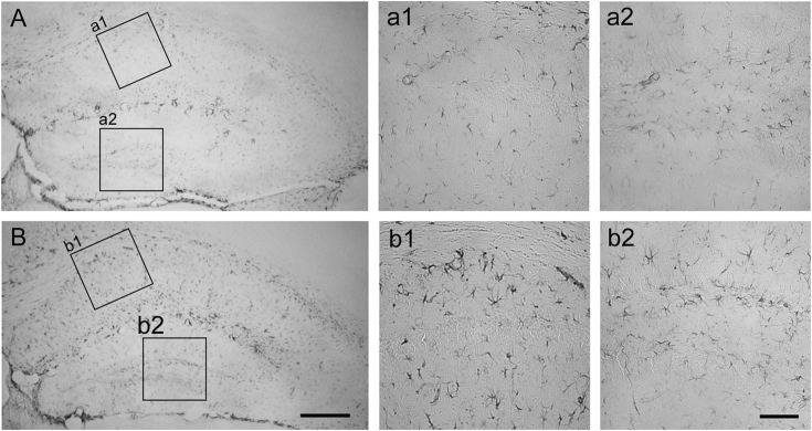
Figure 5Spatial working memory following 4 week-recovery following electromagnetic radiation. (A) The spontaneous alternation score was not significantly difference between two groups suggesting that 4 week long recovery following electromagnetic radiation does not affect spatial working memory. (B) The number of arm entry of exposed-recovered group was significantly higher compared with that of the exposed group. These data suggest that electromagnetic radiation induces hyperactivity-like behavior in mice. Data were collected from 8∼10 mice in each group. n.s. = not significant.
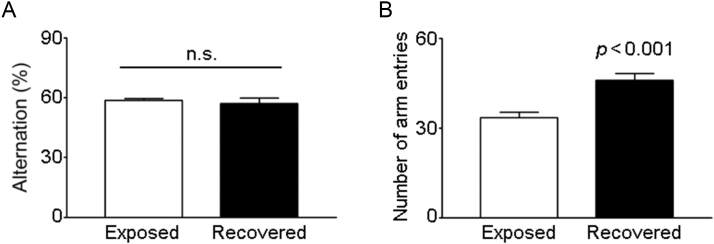
Figure & Data
References
Citations
Citations to this article as recorded by 
- Age-related changes in meningeal lymphatic function are closely associated with vascular endothelial growth factor-C expression
Qi Liu, Cheng Wu, Qian Ding, Xiang-yu Liu, Ni Zhang, Jun-hui Shen, Zi-tong Ou, Tuo Lin, Hong-xiang Zhu, Yue Lan, Guang-qing Xu
Brain Research.2024; 1833: 148868. CrossRef - Education on Electromagnetic Waves Exposure from Smart Devices in Elementary School
Eka Kusumawardhani, Leonardus Sandy Ade Putra, Putranty Widha Nugraheni, Lalak Tarbiyatun Nasyin Maleiva, Romario Aldrian Wicaksono
International Journal of Community Service Learnin.2023; 7(1): 56. CrossRef - Extract of Xylopia aethiopica and its kaurene diterpene, xylopic acid, improve learning and memory in mice
Awo Efua Koomson, Kennedy Kwami Edem Kukuia, Patrick Amoateng, Robert Peter Biney, Thomas Amatey Tagoe, Jeffrey Amoako Mensah, Elvis Ofori Ameyaw, Joseph Torbi, Seth Kwabena Amponsah
IBRO Neuroscience Reports.2022; 12: 249. CrossRef - Functional Differences Between Two Kv1.1 RNA Editing Isoforms: a Comparative Study on Neuronal Overexpression in Mouse Prefrontal Cortex
Liting Zhang, Zetong Peng, Wenjun Bian, Pingping Zhu, Bin Tang, Wei-Ping Liao, Tao Su
Molecular Neurobiology.2021; 58(5): 2046. CrossRef - Electromagnetic shielding properties of cementitious composites containing carbon nanofibers, zinc oxide, and activated carbon powder
Dimuthu Wanasinghe, Farhad Aslani, Guowei Ma
Construction and Building Materials.2021; 285: 122842. CrossRef - A modified four vessel occlusion model of global cerebral ischemia in rats
Wei Sun, Yeting Chen, Yongjie Zhang, Yue Geng, Xiaohang Tang, Runjie Guo, Zean Zhang, Hong Xu, Xuesong Tian
Journal of Neuroscience Methods.2021; 352: 109090. CrossRef - Effects of mild intrauterine hypoperfusion in the second trimester on memory and learning function in rat offspring
Shao-Wei Yin, Yuan Wang, Yi-Lin Meng, Cai-Xia Liu
Neural Regeneration Research.2020; 15(11): 2082. CrossRef - Electromagnetic radiation 2450 MHz exposure causes cognition deficit with mitochondrial dysfunction and activation of intrinsic pathway of apoptosis in rats
Sukesh Kumar Gupta, Manoj Kumar Mesharam, Sairam Krishnamurthy
Journal of Biosciences.2018; 43(2): 263. CrossRef - Age-Dependent Effect of Long-Term Microwave Radiation on Postnatal Neurogenesis in Rats: Morphological and Behavioral Study
A. RAČEK, K. BEŇOVÁ, P. ARNOUL, M. ZÁVODSKÁ, A. ANGELIDIS, V. CIGÁNKOVÁ, V. ŠIMAIOVÁ, E. RAČEKOVÁ
Physiological Research.2018; : 495. CrossRef - Effects of Simulated Mobile Phone Electromagnetic Radiation on Fertilization and Embryo Development
Hong Chen, Zaiqing Qu, Wenhui Liu
Fetal and Pediatric Pathology.2017; 36(2): 123. CrossRef - Effect of rotation preference on spontaneous alternation behavior on Y maze and introduction of a new analytical method, entropy of spontaneous alternation
Jia Bak, Hae-In Pyeon, Jin-I Seok, Yun-Sik Choi
Behavioural Brain Research.2017; 320: 219. CrossRef - Simulation modelling and calculation of dielectric permittivity of Opuntia at 1.7–2.6 GHz
Ediz Delihasanlar, Ahmet Hayrettin Yuzer
Journal of Microwave Power and Electromagnetic Ene.2017; 51(2): 150. CrossRef
 PubReader
 Cite
Cite


